
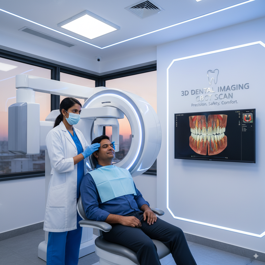
Accurate diagnosis and treatment planning are the foundation of successful dental and surgical care. At 32 Pearls – Dr. Sameer Kaura’s Dental & Maxillofacial Clinic, we use advanced 3D Imaging technology (Cone Beam Computed Tomography – CBCT) to obtain highly detailed views of the teeth, jaws, and facial structures.
Unlike traditional X-rays, which provide only two-dimensional images, CBCT scans capture three-dimensional images that allow for precise evaluation of bone, nerves, and surrounding tissues. This ensures safer, more accurate, and more predictable outcomes in a wide range of dental and surgical procedures.
When is 3D Imaging Used?
-
Dental Implants: To evaluate bone quality, volume, and exact placement sites.
-
Wisdom Tooth Removal: To assess proximity to nerves and sinus cavities.
-
Jaw Surgery & Orthodontics: For precise treatment planning and bite correction.
-
Facial Trauma Cases: To identify fractures and plan reconstructive surgery.
-
Pathology Detection: Early diagnosis of cysts, tumors, and other abnormalities.
Benefits of CBCT 3D Imaging:
-
Provides highly detailed and accurate anatomical views
-
Improves safety by identifying vital structures like nerves and sinuses
-
Allows for more precise implant and surgical planning
-
Reduces risk of complications during surgery
-
Quick, painless, and low-radiation procedure
At 32 Pearls, our state-of-the-art CS 9300 CBCT system provides clear, high-resolution images with minimal radiation exposure, giving both patients and doctors greater confidence in treatment outcomes.
By integrating advanced 3D imaging into every stage of care, we ensure that each procedure — from simple extractions to complex jaw surgeries — is carried out with maximum safety, accuracy, and comfort.